


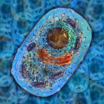
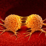
Cell Stress, DNA Damage and Metabolism


Cell stress, DNA damage, and metabolism collectively refer to the cellular response to a diverse range of stressors, including cancer infiltration, infection, extreme temperature fluctuations, exposure to toxins, and mechanical damage. As an oncology contract research organization, we delve into the intricate mechanisms of these cellular processes.
| Cell metabolism (DiHydroFolate Reductase DHFR activity) | Recombinant enzyme |
| Apoptosis assay(Caspase 3/7 activation) | Multiple cellular models |
| Autophagy assay(LC3B turnover assay) | HeLa cells |
| Cell metabolism(Neutral lipids intracellular accumulation) | Multiple cellular models |
| Cell metabolism(Phospholipids intracellular accumulation) | Multiple cellular models |
| Cell metabolism (Protein synthesis quantification) | Multiple cellular models |
| Cell metabolism(Protein synthesis quantification) | Multiple cellular models |
| Cell stress assay (intracellular reactive oxygen species ROS) | Multiple cellular models |
| Cell stress assay (Lipid peroxidation) | Multiple cellular models |
| Cell stress assay (Mitochondrial membrane potential) | Multiple cellular models |
| Hypoxia assay (HIF1α quantification) | Multiple cellular models |
| Hypoxia assay (Pymonidazole metabolite quantification) | Multiple cellular models |
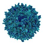
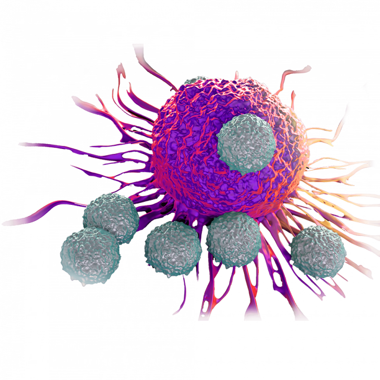
Immuno-oncology
Immuno-oncology, in collaboration with our expertise as an experienced oncology contract research organization, is a specialized field that centers on harnessing the power of the immune system to combat cancer effectively.
| Binding assay of immune check points inhibitors | Multiple inhibitors (HTRF) |
| Immune T-cell infiltration and killing assay cytometry | 3D co-culture multiple cells |
| Immune T-cell killing assay live-content imaging | 2D co-culture multiple cells |
| T-cell activation assay live-content imaging | Human peripheral mononuclear blood cell (hPBMC/CD3 cells) |
| Colon cancer (anti-PD-1/CTLA-4) | CT26.WT cells Mouse |
| Glioblastoma (anti-PD-1/CTLA-4) | GL261 cells Mouse |
| Renal cancer (anti-PD-1/CTLA-4) | RenCa cells Mouse |
Receptor Pharmacology & Signaling Pathways
Receptor pharmacology and signaling pathways, a fundamental aspect of cancer research, involves studying the receptors and molecular pathways that play a pivotal role in controlling cancer growth and progression.
| AKT phosphorylation | Multiple cellular models |
| Androgen receptor nuclear translocation | LNCaP cell line |
| Calcium homeostasis | Multiple cellular models |
| cAMP quantification | Multiple cellular models |
| ERK activation (pERK1/2) | Multiple cellular models |
| NFkB activation | Multiple cellular models |
| Prostate Specific Antigen (PSA) expression | LNCap cell line |
Targeting Angiogenis
As a recognized oncology contract research organization, Porsolt focuses on targeting angiogenesis, a critical strategy in cancer research. Angiogenesis refers to the formation of new blood vessels that nourish tumors.
| Assay (screening – 3R approach) | Chicken eggs |
| Tube formation assay live-content imaging | HUVECs cells co-cultured with fibroblasts |
Targeting Metastasis
Metastasis occurs when cancer cells spread from their primary site of origin to other areas of the body. By developing innovative strategies to target and inhibit this process, our oncology contract research organization capabilities aim to improve cancer treatment outcomes and patient safety and survival.
| Experimental metastasis CAM Assay (screening – 3R approach) | Chicken eggs |
| Experimental lung metastasis syngeneic model of breast cancer | 4T1 cells |
| Experimental lung metastasis syngeneic model of colon cancer | CT26.WT cells |
| Experimental lung metastasis xenograft model of breast cancer | MDA-MB-231 cells Mouse |
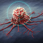
Targeting Tumor Cells
As a recognized oncology contract research organization, Porsolt specializes in targeting specific tumor cells as a crucial approach to combat cancer.
| Chicken Chorioallantoic Membrane (CAM) xenograft assay (screening – 3R approach) | Multiple cellular models Chicken eggs |
| Apoptosis assay | Multiple 2D cellular models |
| Cell cycle cytometry analysis | Multiple 2D or 3D cellular models |
| Cell proliferation/cytolysis assay high-content imaging | Multiple 2D cellular models |
| Cell viability colorimetric assay | Multiple 2D cellular models |
| Clonogenicity assay (anchorage-independent) | Multiple 3D cellular models |
| Invasion assay | Multiple 3D cellular models |
| Migration assay – high-content imaging | Multiple 2D cellular models |
| Spheroid proliferation/cytolysis assay high-content imaging | Multiple 3D cellular models |
| Hollow fiber assay Multiple cellular models (screening – 3R approach) | Mouse – Rat |
Orthotopic syngeneic models of:
| Breast cancer | 4T1 cells – Mouse |
| Colon cancer including neurological and behavior analysis | CT26.WT/C26 cells - Mouse |
| Glioblastoma (brain tumor) | GL261 cells - Mouse |
| Kidney cancer | RenCa cells - Mouse |
| Lung cancer including respiratory function | LLC1/KLN205 cells - Mouse |
Orthotopic xenograft models of:
| Breast cancer | MDA-MB -231 cells - Mouse |
| Lung cancer | A549/PC-9 cells Mouse |
| Glioblastoma | U87MG cells in mouse |
Subcutaneous syngeneic model of:
| Breast cancer | 4T1 cells – Mouse |
| Colon cancer | CT26.WT/C26 cells - Mouse |
| Glioblastoma (brain cancer) | GL261 cells - Mouse |
| Lung cancer | LLC1/KLN205 cells - Mouse |
| Renal Cancer | 105K cells (Tuberous Sclerosis Alliance) |
Subcutaneous xenograft models of:
| Bladder cancer | SW780 cells - Mouse |
| Breast cancer | MDA-MB -231/BT-20 cells Mouse |
| Colon cancer | HCT-8/HCT-116 cells Mouse |
| Fibrosarcoma | HT-1080 cells - Mouse |
| Glioblastoma (brain tumor) | U118MG/U87MG/U138MG cells Mouse |
| Kidney cancer | ACHN cells - Mouse |
| Liver cancer | Hep3B2.1-7/HepG2 cells Mouse |
| Lung cancer | A549/PC-9 cells - Mouse |
| Pancreatic cancer | PANC-1 cells - Mouse |
| Prostate cancer | LNCaP/PC-3 - Mouse |

Tumor-Associated Side Effects
Our oncology contract research organization investigates tumor-associated side effects, which are secondary reactions arising from cancer treatments.
PAIN
IN VIVO MODELS| Chemotherapy-induced intestinal mucositis | DRGs Rat |
| Chemotherapy-induced intestinal mucositis | Mouse |
| Chemotherapy - induced neuropathic pain: Vincristine model | Rat |
CACHEXIA
IN VIVO MODELS| Drug-induced cachexia model | Rat |
| Tumor-induced cachexia model | AH-130 cells (Rat) |
| Tumor-induced cachexia model | C26 cells (Mouse) |
| Tumor-induced cachexia model | LLC1 cells (Mouse) |

Any questions or concerns ?
CONTACT US
